SignalSCREEN
an automated high-throughput confocal imaging platform
SignalSCREEN is designed to analyze seedlings of different plant species for disease resistance and susceptibility phenotypes. Seedlings are analyzed in multi-well plates that are automatically loaded onto an inverse microscope and analyzed by live cell imaging (Figure 1). The spinning disc microscope allows us to image all standard fluorophores that are on the market today. Plant-pathogen interactions are analyzed in roots and in aerial tissues. For the analysis of aerial tissues, the seedlings are embedded in top agar and the plates are flipped in an inverse position. SignalSCREEN is used to quantify the propagation of fluorescently labelled pathogens by live cell/tissue imaging of large collections of seedlings. Additionally, the platform can be used for invasive screens, in which the accumulation of fluorescently stained defense markers is quantified in plant tissue fragments (Figure 1).
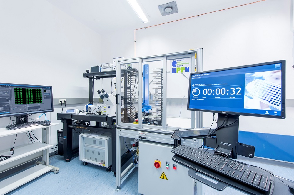
Figure 1: Platform for automated high-throughput confocal imaging
Our growing pathogen collection includes 38 bacterial pathogens, of which 22 have been marked with GREEN FLUORESCENT PROTEIN (GFP). Together with more than 100 fungal pathogens, this collection can be used to infect a growing number of plant species, including tomato, potato, tobacco, the model dicotyledonous plant Arabidopsis thaliana, and the monocots barley, wheat, and maize. We analyze, for example, Arabidopsis thaliana collections in 96-well plates for the susceptibility of the aerial tissues to GFP-marked bacterial pathogens (Figure 2). Wells are screened with 10X or 20X objectives and images are recorded in one of three patterns (3X3, 4X4, or doughnut). The wells from each run (that can include up to 44 multi-well plates) are automatically ranked by fluorescens intensity.
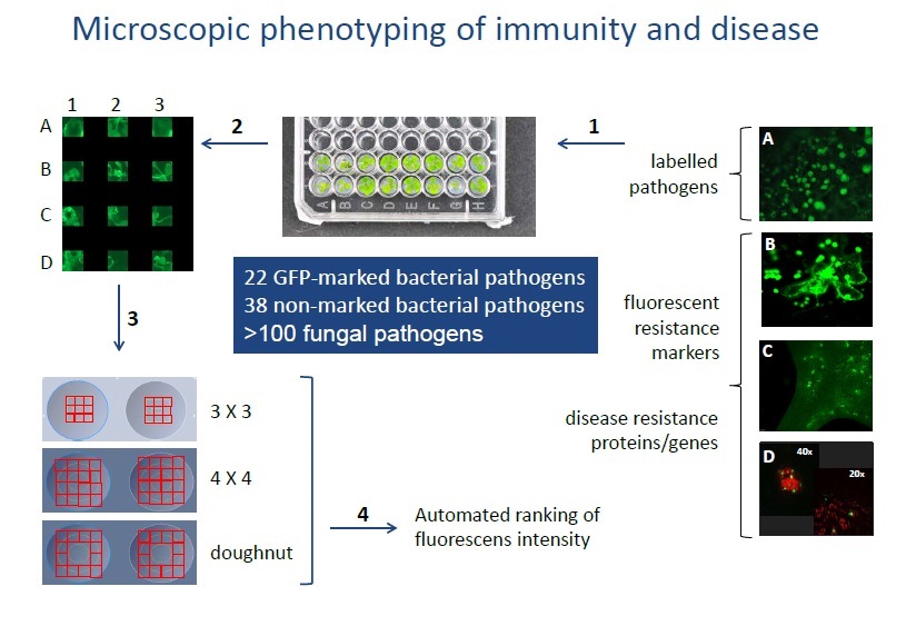
Figure 2
Immediately following the analysis, phenotypes-of-interest can be isolated and the corresponding seedlings grown for further analysis and/or for seed propagation. Further analysis can include imaging applications at high speed or at higher magnification in SignalSCREEN (40X and 63X) or at high resolution using a stand-alone confocal laser scanning microscope (coming to us soon).